
Search Your Topic
Glycosaminoglycans and proteoglycans – A quick revision
Introduction
- The most abundant heteropolysaccharides in the body.
- Highly negatively charged molecules, with extended conformation that imparts high viscosity to the solution.
- GAGs are located primarily on the surface of cells or in the extracellular matrix (ECM).
- Along with the high viscosity of GAGs comes low compressibility, which makes these molecules ideal for a lubricating fluid in the joints.
- Their rigidity provides structural integrity to cells and provides passageways between cells, allowing for cell migration.
GAGs of physiological significance
The specific GAGs of physiological significance are:
- Hyaluronic acid,
- Dermatan sulfate
- Chondroitin sulfate
- Heparin
- Heparan sulfate, and
- Keratan sulfate.
Chemistry
- These molecules are long unbranched polysaccharides containing a repeating disaccharide unit. [acidic sugar-amino sugar]n
- Although each of these GAGs has a predominant disaccharide component, heterogeneity does exist in the sugars present in the make-up of any given class of GAG.
Nature of amino sugars (figure-1)
The disaccharide units contain either of two modified amino sugars,
- N-acetyl galactosamine (GalNAc) or
- N-acetylglucosamine (GlcNAc),

Figure-1- Amino sugars- β-D Glucosamine and β-D Galactosamine
The amino sugar may also be sulfated on carbon 4 or 6 or on non-acetylated nitrogen.
Nature of acid sugar
Uronic acid represents acid sugar in the form of:
- Glucuronate or
- Iduronate
The acidic sugars contain carboxyl groups that are negatively charged at physiological pH, (figure-2) and together with the sulfate groups, give glycosaminoglycans their strongly negative nature.

Figure-2- The acid sugars present in Glycosaminoglycans are D- Glucuronate, and L- Iduronate.
Structure-function relationship
Because of their large number of negative charges, these heteropolysaccharides chains tend to be extended in solution. They repel each other and are surrounded by a shell of water molecules. When brought together they “slip” past each other. This produces the slippery consistency of mucous secretions and synovial fluid. When a solution of GAG is compressed, the water is squeezed out and GAGs are forced to occupy a smaller volume. When the compression is released the GAGs get back to their original, hydrated volume because of the repulsion of the negative charges. This property contributes to the resilience of the synovial fluid and vitreous humor of the eye.
THE SPECIFIC GAGs OF PHYSIOLOGICAL SIGNIFICANCE ARE:
1) Hyaluronic acid – The repeating disaccharide unit is:
Glucuronic acid and N Acetylglucosamine (figure-3)
(D-Glucuronate + GlcNAc) n

Figure-3- The structure of hyaluronic acid
Occurrence: Hyaluronic acid is found in –
- Synovial fluid,
- ECM of loose connective tissue, umbilical cord and vitreous humor of the eye.
Function
- It serves as a lubricant and shock absorber.
- It is the only GAG that is not limited to animal tissue but is also found in bacteria.
- Hyaluronic acid is unique among the GAGs because it does not contain any sulfate and is not found covalently attached to proteins.
- It forms non-covalently linked complexes with Proteoglycans in the ECM.
- Hyaluronic acid polymers are very large (100 – 10,000 k Da) and can displace a large volume of water.
2) Dermatan sulfate- The repeating disaccharide unit is L-Iduronic acid and N-Acetyl Galactosamine with a variable amount of Glucuronic acids (figure-4).
(L-Iduronate + GalNAc sulfate) n

Figure-4- The structure of Dermatan Sulfate
Occurrence: It is found in skin, blood vessels, and heart valves
3) Chondroitin sulfate- The repeating disaccharide unit is Glucuronic acid and N-Acetyl galactosamine with sulfate on either C-4 or C-6. Based on the presence of the sulfate group, it may be labeled as Chondroitin-4-Sulfate or Chondroitin-6-Sulfate (figure-5).
(D-Glucuronate + GalNAc sulfate) n

Figure-5- The structure of Chondroitin Sulfate
Occurrence: It is found in cartilages, tendons, ligaments, heart valves and aorta.
Function
It is the most abundant GAG. In cartilages, it binds collagen and holds fibers in a tight, strong network.
4) Heparin sulfate – The repeating disaccharide unit is:
L-Iduronic acid and D- Glucosamine with variable amounts of Glucuronic acid. Most glucosamine residues are bound in Sulfamide linkages (figure-6). Sulfate is also found on C-3 or C-6 of Glucosamine and C-2 of uronic acid (An average of 2.5 Sulfate per disaccharide unit)
(D-Glucuronate sulfate +N-Sulfo-D-glucosamine) n

Figure-6- The structure of Heparin Sulfate
Occurrence: Heparin is a component of intracellular granules of mast cells lining the arteries of the lungs, liver, and skin (contrary to other GAGs that are extracellular compounds, it is intracellular).
Function– It serves as an anticoagulant.
5) Heparan sulfate: Heparans have fewer sulfate groups than heparins. The repeating disaccharide unit is the same as Heparin. Some Glucosamines are acetylated
Occurrence- It is an extracellular GAG found in the basement membrane and as a ubiquitous component of cell surfaces
6) Keratan sulfate –The repeating disaccharide unit is galactose and N-Acetyl glucosamine (No uronic acid). The sulfate content is variable and may be present on C-6 of either sugar (figure-7).
(Gal + GlcNAc sulfate) n

Figure-7- The structure of Keratan sulfate
Occurrence: cornea, bone, cartilage; Keratan sulfates are often aggregated with Chondroitin sulfates.
Proteoglycans (mucoproteins)
Proteoglycans are formed of glycosaminoglycans (GAGs) covalently attached to the core proteins. They are found in all connective tissues, extracellular matrix (ECM) and on the surfaces of many cell types. Proteoglycans are remarkable for their diversity (different cores, different numbers of GAGs with various lengths and compositions).
Structure of Proteoglycans
All of the GAGs, except Hyaluronic acid, are found covalently attached to protein forming proteoglycan monomers.
Structure of Proteoglycan monomer
A Proteoglycan monomer found in cartilage consists of a core protein to which the linear GAG chains are covalently linked. These chains which each may be composed of more than 100 monosaccharides extend out from the core protein and remain separated from each other because of charge repulsion. The resulting structure resembles a ‘Bottlebrush’ (figure-8). In cartilage proteoglycans, the species of glycosaminoglycans include Chondroitin sulfate and Keratan sulfate.

Figure-8- The structure of Proteoglycan monomer (Bottle Brush)
The Linkage between the carbohydrate chain and the protein
The linkage of GAGs such as (heparan sulfates and Chondroitin sulfates) to the protein core involves a specific trisaccharide linker (Galactose-galactose-Xylose). The protein cores of Proteoglycans are rich in Serine and Threonine residues which allow multiple GAG attachments.
An O-Glycosidic bond is formed between the Xylose and the hydroxyl group of Serine. Some forms of Keratan sulfates are linked to the protein core through an N-asparaginyl bond (N- Glycosidic linkage)
Proteoglycan Aggregates- The proteoglycan monomers associate with a molecule of Hyaluronic acid to form Proteoglycan aggregates (figure-9). The association is not covalent but occurs primarily through ionic interactions between the core protein and Hyaluronic acid. The association is stabilized by additional small proteins called Link proteins.
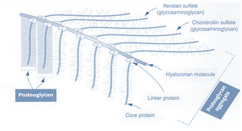
Figure-9- The structure of proteoglycan aggregate
Functions of Proteoglycans
They perform numerous vital functions within the body.
GAG dependent functions can be divided into two classes: the biophysical and the biochemical.
1) The biophysical functions depend on the unique properties of GAGs: the ability to fill the space, bind and organize water molecules and repel negatively charged molecules. Because of high viscosity and low compressibility they are ideal for a lubricating fluid in the joints. On the other hand, their rigidity provides structural integrity to the cells and allows cell migration due to providing the passageways between cells.
2) The other, more biochemical functions of GAGs are mediated by specific binding of GAGs to other macromolecules, mostly proteins. Proteoglycans participate in cell and tissue development and physiology.
3) Heparin acts as an anticoagulant and is used in clinical practice.